Using advanced microscopy to examine cell structure and function
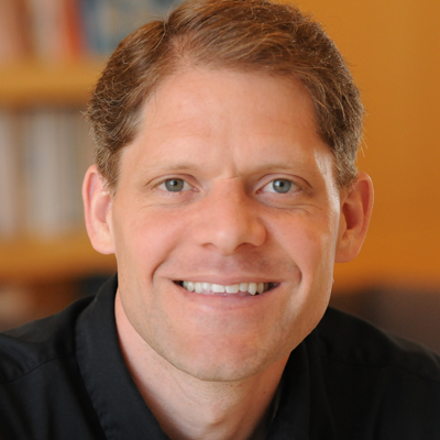
Some bacteria assemble long needles on their exterior that "poke" into our cells, injecting toxins, while others have harpoon-like spikes that fly out of the bacteria and puncture cells around them. Some sporulate to protect themselves, and others hide in biofilms, but how all this works is not yet understood. Dr. Grant Jensen, Professor of Biophysics and Biology at California Institute of Technology, uses the most advanced electron microscopes available today to image the internal machinery inside cells and see how it all fits together. It is important to learn about all cells and viruses can do so we can improve our abilities to combat the dangerous ones and exploit the others. Eventually we might even be able to mix and match parts to create new forms of cellular life that desalinate water, produce clean energy, clean the environment, or clear infections.
Dr. Jensen’s lab of 15 professional and training researchers have the world’s best electron microscope for probing the interiors of cells and viruses. Unlike traditional light microscopes that are limited by the wavelength of light (and therefore only allow one to see small bacterial cells but not what’s inside them), electron microscopes can probe objects within the cells. The team particularly uses and helps develop cryo-electron microscopy, which preserves cells in a life-like, frozen state, unstained. Producing 3D images of cells much like a CAT scan of one’s brain, Dr. Jensen’s team is at the forefront of visualizing the internal details of cells and viruses, addressing fundamental questions regarding the movement and behavior of bacteria and bacterial infections.
Current projects include:
- Enhancing Microscopy Techniques: Dr. Jensen and his team are constantly working to develop microscopy techniques that will enable seeing inside cells and viruses in ways that were previously impossible. They work with instrument companies to test new developments such as high-end cameras or new sample holders. The team is also working on methods to accelerate data collection and store and organize the many tens of terabytes of image data they produce. They build software and tools to interrogate images and have established a system to correlate light microscope images with electron microscope images to identify and characterize machines present inside cells.
- Understanding Cell Machinery: Dr. Jensen’s advanced microscopy techniques allow his team to study how cells maintain certain shapes, know where to swim, and stick to surfaces like human cells. For example, Dr. Jensen’s team has discovered that many bacteria have “spring-loaded molecular daggers,” or harpoon-like tubes inside of them, and when they bump into prey cells, the tube contracts and shoots the javelin through the prey cell. Ultimately, Dr. Jensen wants to understand at a molecular level how cells do what they do.
- Understanding HIV: Dr. Jensen is applying his imaging techniques to understand the HIV virus and how it infects human cells, spreading and causing disease. His lab was the first to produce 3D images of the HIV virus, visualizing the viruses in both their immature state and their mature state. The genome of HIV that ultimately gets inserted into a human chromosome is protected and carried within a protein shell called capsid, and Dr. Jensen’s work has produced a more complete picture of how the protein capsid assembles and disassembles and how certain cells manage to resist infection.
Bio
For the past 12 years, Dr. Grant Jensen has led a large research lab of 10-20 people at the California Institute of Technology studying the structure and function of large macromolecular complexes. His team specializes in electron cryotomography (ECT), the highest resolution technique available today to image unique objects such as intact cells and viruses.
Dr. Jensen grew up in Los Alamos, NM, birthplace of the nation’s nuclear technologies. Los Alamos county boasted the highest number of Ph.D.s per capita of any county in the entire US; it seemed like nearly everybody in the town was a physicist. For a kid growing up in that environment who did well in school, everyone automatically assumed he would grow up to be a physicist! In fact, when Dr. Jensen was a senior in high school, there was a large group of students wanting to take a college-level physics class. Unfortunately, the teacher who was supposed to teach the course suddenly fell sick, and the school was not able to offer the class any longer. So, several fathers, including Dr. Jensen’s own, volunteered to teach the class.
The year after Dr. Jensen graduated from high school, the Soviet Union collapsed, and there was a feeling throughout the little town that science had saved our country's liberty through the Cold War. Science was what mattered. Science moved boundaries -- it changed the world -- other professions mostly just maintain it. “When viewed from a distance,” Dr. Jensen remarks, “the history of mankind is not punctuated by pharaohs and emperors and presidents or even world wars so much as science: the domestication of animals, the development of irrigation, the discovery of bronze and then iron and then steel, vaccines and antibiotics, internal combustion engines, fertilizer and electricity. So of course I chose science!” He chose electron microscopy because it is the most direct way to learn about how cells and viruses actually work -- when we can "see" them in action at a molecular level, much of how they work becomes clear.
The most “bizarre” thing about Dr. Jensen is that he has six children, the eldest one being 20 years old. His “hobby”, outside of research, is playing with his children and attending their activities.
For more information, visit http://www.jensenlab.caltech.edu/
Publications
Videos
Awards
Organizer, World Congress on Electron Tomography, 2014
Member of the Scientific Advisory Board, 2010 - present
Boulder Laboratory for 3-D Electron Microscopy of Cells
Member of the Defense Science Study Group, 2010 - 2011
Searle Scholar, 2004 - 2006
Damon Runyon-Walter Winchell postdoctoral fellow, 1999 - 2002
Patents
U.S. Patent No. 6,604,052: “Methods and compositions for use in three-dimensional structural determination.”
Jensen, G. J., and Kornberg, R. D. (1999). August 5, 2003.


