Breakthrough neuroimaging helps understand healthy and abnormal brains
In brain sciences perhaps the single most important conceptual revolution, now underway, is a view of the brain as a set of nested, hierarchical and interacting networks. It is not just local modules that are specialized for particular functions. These exist, but they sit on top of a much richer network architecture that scientists are now finally able to map with state-of-the-art human brain mapping methods. Dr. Scott Grafton, of the University of California, Santa Barbara, has focused on developing better methods to measure structural networks in the brain. These are the connections that link different modules together. While many try to do this, most methods are quick, noisy, and incomplete. Dr. Grafton and his team can generate dense, thorough and reproducible maps of brain connections. He develops software tools to visualize and quantify these connections. By breaking down dogma about how the brain ought to work locally, Dr. Grafton's research is beginning to show how it globally operates. This network based understanding provides practical approaches that are applicable to understanding individuality, and accelerating learning in healthy people. In addition, his research is helping to better understand obsessive compulsive disorder, depression, developmental disorders, Parkinson's disease, head injury and more.
While many labs use brain scanning resources, Dr. Grafton's is unique because he and his team combine state-of-the-art imaging, sophisticated analysis abilities, a development environment for building new analysis tools, and a focus on research questions that are rooted in fundamental clinical issues. At UCSB, he has freedom from the yoke of traditional medical school environments, and an excellent talent pool to draw from. Such a combination lends to sophisticated collaboration among lab members in varying disciplines and outside the medical realm. The overarching questions of Dr. Grafton's lab focused upon understanding the fundamental mapping of the brain in addition to the translational application of this work for patients with brain injuries, strokes, or developmental disorders. At an exciting stage now, Dr. Grafton's research is shifting from the hard work of optimizing imaging methods to the collection of large clinical datasets as well as the creation of novel image analysis tools. Therefore, in the near future, his team hopes to create the best data sets available for a target set of clinical disorders and to release a set of software tools for analyzing brain connectivity.
Dr. Grafton's methods and tools have a number of potential applications including:
-
Mild traumatic brain injury: Roughly 1 in 10 people who get a concussion do not recover. Dr. Grafton and his team are using their methods to identify those with "broken" connections with great precision thereby helping to treat those that do not recover.
-
Developmental disorders: Dr. Grafton is interested in learning why disabilities like stutters or learning difficulties appear in people that are otherwise normal. He and his team are using their methods to demonstrate abnormalities of brain connections that probably arise during development of the fetus. Most importantly, Dr. Grafton wants to know if the wiring is different for those who recover or respond to therapy and those that don't.
-
Individual differences: Why are some people good at chess. Others at playing the piano? Does wiring matter? Dr. Grafton hopes to answer such questions by mapping out the wiring of the human brain and thus, help to shed light upon individual differences.
-
Rewiring: There is growing evidence that when we learn, we adjust the speed with which brain connections transmit information. This is radically different from what is done in computers, where conductions in the wires are all constant. Dr. Grafton is using his method to test this radical concept.
-
Structure vs. Function: Dr. Grafton and his team can measure brain activity as people learn and perform motor skills and ask how this activity relates to underlying wiring. In collaboration with mathematicians, computer scientists and complex dynamics physicists he and his team develop tools to assess these relationships.
Bio
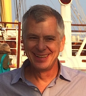
Dr. Grafton has been interested in understanding the large scale, macroscopic organization of the human brain since college, when he worked as a researcher at the National Institutes of Mental Health. While there are many investigators considering the molecules and cellular activities of the brain, for him it is far more interesting to understand the overall design features. With his training in neurology, he was an early developer of methods for functional brain mapping using PET scanning and then functional MRI. As a clinical neurologist with 20 years of patient experience, he used imaging to understand why patients with disorders like Parkinson's disease can't move normally. He used such methods to determine why new medicines, surgery, and deep simulators led to improvements. When Dr. Grafton moved to Dartmouth College, he opened the first free-standing MRI brain imaging center in the US that was not under the supervision of radiologists or a medical school. He thus demonstrated that this research paradigm could work and that cognitive neuroscientists could work directly with MRI to study the brain.
In his free time, aside from research, Dr. Grafton's love of mapping is extended beyond the brain and to the wilderness. He enjoys backcountry and wilderness hiking as well as off-trail hiking and running. In addition, he loves to fly airplanes.